

SHAH ALAM, Nov 4 — For most people, worsening eyesight means getting a stronger pair of glasses. But when spectacle prescriptions keep changing and vision remains blurred even with new lenses, the problem may lie deeper than short-sightedness or astigmatism.
It could be keratoconus, a progressive condition in which the cornea – the transparent surface at the front of the eye – gradually thins and bulges into a cone, distorting the way light enters the eye and impairing vision.
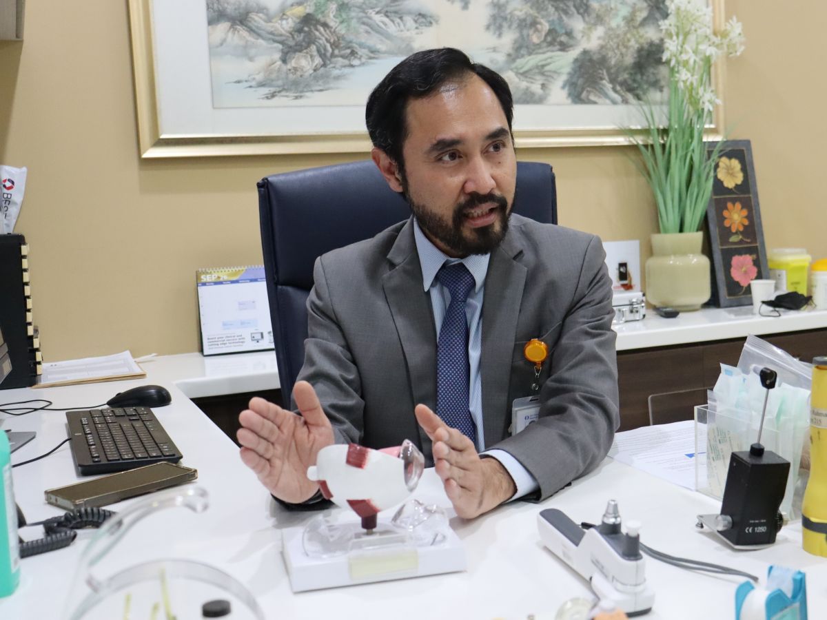
“If you find yourself visiting the optical shop frequently and still cannot see clearly enough with your glasses, please see your eye doctor,” Dr Ainur Rahman Anuar Masduki, consultant ophthalmologist and corneal surgeon at Ara Damansara Medical Centre (ADMC), told CodeBlue in an interview.
Keratoconus often goes undiagnosed because its early symptoms can resemble ordinary refractive errors. But as the condition progresses, the cornea becomes increasingly irregular, and glasses may no longer adequately correct the resulting visual distortion.
“Some people have keratoconus but don’t know that they have it. They remain undiagnosed and, unfortunately, some progress to the later stages, when their vision becomes severely impaired and they may need a corneal transplant,” Dr Ainur said.
Keratoconus Often Develops In Young People

Keratoconus commonly develops during adolescence or early adulthood, with the condition potentially progressing faster in children. It usually affects both eyes, although one may be more severely affected.
There is no single known cause of keratoconus. Its development is associated with genetic factors and family history, as well as conditions such as asthma, eczema, and allergies. Frequent or vigorous eye rubbing is also a recognised risk factor.
Parents should be alert to children and teenagers who frequently rub their eyes, particularly those with allergies.
One early warning sign is a spectacle prescription that keeps changing, particularly increasing astigmatism. Patients may also experience blurred or distorted vision, ghost images, glare, halos around lights, and difficulty driving at night. As the condition progresses, even new glasses may no longer provide clear sight.
Reliable data on keratoconus prevalence in Malaysia is lacking, Dr Ainur said, although international studies have reported relatively higher rates in Asian and Middle Eastern populations.
Early keratoconus can be difficult to detect during a standard spectacle prescription test at an optical shop, which checks for changes in short-sightedness and astigmatism.
Identifying abnormalities in the cornea’s shape and thickness requires specialised imaging. Ophthalmologists use corneal tomography, with devices such as the Pentacam, to map the cornea and detect changes that may otherwise go unnoticed.
Although not every case can be prevented, avoiding eye rubbing and managing allergies can help address associated risk factors.
People whose spectacle prescriptions change rapidly, or who continue to experience blurred vision despite new glasses, should seek an assessment by an ophthalmologist.
Treatment depends on the severity of the condition and whether it is progressing. In earlier stages, spectacles and soft contact lenses may be sufficient to correct vision, Dr Ainur said.
As the cornea becomes more irregular, patients may need rigid contact lenses, which can be uncomfortable and difficult to tolerate.
For patients whose condition is progressing, corneal collagen cross-linking can help strengthen the cornea and slow or stop further deterioration.
The procedure involves applying riboflavin (vitamin B2) drops to the cornea before exposing it to ultraviolet light. The light activates the riboflavin, triggering a process that strengthens the bonds between collagen fibres in the cornea.
This makes the cornea stiffer and helps slow or stop further progression of the condition, but does not reverse existing deformation or restore vision already lost.
In advanced cases, a corneal transplant may be necessary. The operation replaces damaged corneal tissue, but recovery can be prolonged and complications include graft rejection, graft failure, and glaucoma.
CAIRS Offers Another Option For Selected Patients

For selected patients with more advanced keratoconus, a newer procedure offers another option to improve vision by adding donor tissue to the cornea rather than removing the patient’s own tissue.
Known as Corneal Allogenic Intrastromal Ring Segments surgery, or CAIRS, the procedure uses small segments of processed donor corneal tissue to reshape the patient’s cornea.
A specialised femtosecond laser, which makes precise cuts within the cornea, creates a narrow tunnel through which the surgeon inserts the curved donor tissue segments.
These implants help flatten the cone-shaped bulge and make the cornea more regular, potentially improving vision.
“When you do CAIRS, you don’t remove any part of the patient’s cornea. We in fact insert or add tissue to the cornea, rather than remove their natural cornea,” said Dr Ainur.
The implants are made from human corneal tissue donated by deceased donors. Unlike a conventional corneal transplant, CAIRS uses tissue that has been processed into small segments by a specialised company, allowing it to be stored for about two to three years.
This means surgeons do not have to wait for a fresh donor cornea to become available before performing the procedure.
Dr Ainur said the tissue is also processed to reduce its antigenicity, making an immune response less likely.
No genetic or tissue matching is required, while the number, size, and placement of the segments are tailored to each patient’s corneal shape and the position and severity of the cone.

ADMC performed its first CAIRS procedure in July 2024, which Dr Ainur described as the first reported use of the technique in Malaysia and Southeast Asia. The hospital has since performed about 35 cases.
Dr Ainur said CAIRS may benefit patients who can no longer achieve satisfactory vision with spectacles or contact lenses, particularly those who depend on rigid lenses that can be uncomfortable and difficult to tolerate.
Some patients notice an improvement as early as the day after surgery, while others take six months to a year to see changes as the cornea gradually remodels.
The aim is to make the cornea more regular and vision easier to correct, rather than necessarily making patients spectacle-free. Some may still need ordinary glasses or specialised contact lenses.
Unlike LASIK, which reshapes the cornea by removing tissue, CAIRS adds donor tissue. It also differs from a corneal transplant, which replaces damaged corneal tissue.
Because keratoconus can continue to progress, patients require long-term monitoring. Some may eventually need corneal cross-linking to stabilise the cornea, as CAIRS reshapes it but does not necessarily stop the underlying condition from progressing.
Recovery is generally less involved than after a full corneal transplant. CAIRS does not require the stitches used to secure a transplanted cornea. Patients typically use antibiotic drops for a week and steroid drops for about a month, alongside lubricating drops.
This is a shorter steroid course than after a full corneal transplant, when patients may need steroid drops for an extended period, sometimes for life, to reduce the risk of rejection. Steroids help suppress inflammation and the immune response against transplanted tissue.
At ADMC, CAIRS costs approximately RM15,000 per eye, according to Dr Ainur. He said all his patients so far had received insurance coverage, although this depends on individual policy terms and patients should check with their insurers before proceeding.


